Full Name
IKK epsilon (701 - 716)
Long Name Textual
inhibitory kB kinase epsilon
Immuno Sequence
NRIIERLNRVPAPPDV [residues 701 - 716 of human]
Mono/Poly clonal
Polyclonal
Gel Image

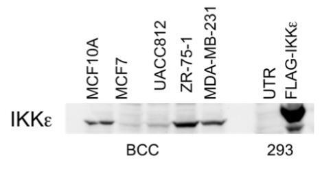
25 ug of cell lysates from various breast cancer cell lines and HEK293 cells transfected with either empty vector (UTR) or FLAG tagged IKK epsilon were separated on an 8 % SDS-Page gel and transferred to PDVF membrane. The membrane was immunoblotted with anti-IKK epsilon (701 – 716) at 1 ug/ml. Binding of the primary antibody was detected using rabbit peroxidase conjugated anti-sheep IgG antibody (1 in 10, 000 dilution, Pierce) followed by enhanced chemiluminescence (ECL, Amersham).
![25 ug of HEK293 cells transfected with either empty vector (control), FLAG tagged TBK1 or FLAG tagged IKK epsilon were separated on an 8 % SDS-Page gel and transferred to PDVF membrane. The membrane was immunoblotted with anti-IKK epsilon (701 – 716) at 1 ug/ml (upper panel) or anti-FLAG M2 [Sigma] (lower panel). Binding of the primary antibody was detected using rabbit peroxidase conjugated anti- sheep IgG antibody (1 in 10, 000 dilution, Pierce) followed by enhanced chemiluminescence (ECL, Amersham).](/sites/default/files/styles/gel_image/public/gel_images/IKK-epsilon_701-716_S255C_IP.jpg?itok=R9z27ikk)
25 ug of HEK293 cells transfected with either empty vector (control), FLAG tagged TBK1 or FLAG tagged IKK epsilon were separated on an 8 % SDS-Page gel and transferred to PDVF membrane. The membrane was immunoblotted with anti-IKK epsilon (701 – 716) at 1 ug/ml (upper panel) or anti-FLAG M2 [Sigma] (lower panel). Binding of the primary antibody was detected using rabbit peroxidase conjugated anti- sheep IgG antibody (1 in 10, 000 dilution, Pierce) followed by enhanced chemiluminescence (ECL, Amersham).

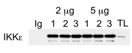
1 mg of protein extract from RAW264.7 macrophages was incubated with pre-immune IgG (Ig) or either the first, second or third bleeds of affinity purified anti-IKK epsilon (701–716)at2or5ug. Thesampleswereincubatedfor1hrat4degConanendover end roller. 10 ul of Protein G Sepharose was added and incubated for a further 15 mins at 4 deg C. The beads were collected by centrifugation and washed three times in lysis buffer. The immunoprecipitates were separated on an 8 % SDS-Page gel, along with 20 ug of total protein extract (TL), and transferred to PDVF membrane. The membrane was immunoblotted with anti-IKK epsilon (Sigma) followed by HRP-conjugated anti-mouse antibodies with detection by enhanced chemiluminescence (ECL, Amersham).
PDF Datasheet
IKK epsilon373.73 KB
Sheep No
S255C
Unit Source
Aliquot
0.1mg
Price
£150.00


